Glaucoma Center of Michigan
Glaucoma Center of Michigan
Glaucoma Suspect and Ocular Hypertension are terms used to describe conditions where a patient does not currently have glaucoma, but is at a higher risk of developing it in the future. Because glaucoma is the “sneak thief of sight,” identifying these conditions early is the best way to prevent permanent vision loss.
Being told you are a “glaucoma suspect” does not mean you have the disease. Rather, it means your eye doctor has found one or more clinical findings that are often seen in people with glaucoma. Think of it like having “pre-hypertension” or “pre-diabetes”; it is a warning sign that requires closer monitoring to ensure your eye health stays stable.
You may be considered a glaucoma suspect if:
Ocular hypertension is a specific condition where the fluid pressure inside your eye (intraocular pressure) is higher than normal, but the optic nerve remains healthy and there is no loss of peripheral vision.
To understand this, remember how the eye works: the eye is filled with a fluid called aqueous humor. This fluid is constantly produced and drained to maintain a healthy pressure. If the “drainage canal” becomes slightly sluggish or the eye produces too much fluid, the pressure rises—much like a balloon that is slightly overfilled with air.
While not everyone with high eye pressure will develop glaucoma, those with ocular hypertension are at an increased risk. Because high pressure itself causes no pain or symptoms, it can only be detected through a professional eye examination.
We monitor suspects and those with ocular hypertension closely because certain factors increase the likelihood of these conditions turning into glaucoma:
Because vision lost to glaucoma is irreversible, our goal at the Glaucoma Center of Michigan is prevention. If you are a suspect or have ocular hypertension, we may not start treatment (such as eye drops) immediately. Instead, we perform regular “baseline” testing to see if your eyes change over time.
If we detect the very first signs of damage to the optic nerve or a change in your side vision, we can begin treatment immediately to stop the disease in its tracks.
To track your eye health, we use the same advanced diagnostic tools used to detect glaucoma:
Observation of the drainage angle is performed by using a gonioscope, a specially mirrored and magnified lens that functions like a periscope.
The lens magnifies and bends light to allow observation of the structures of the trabecular meshwork.
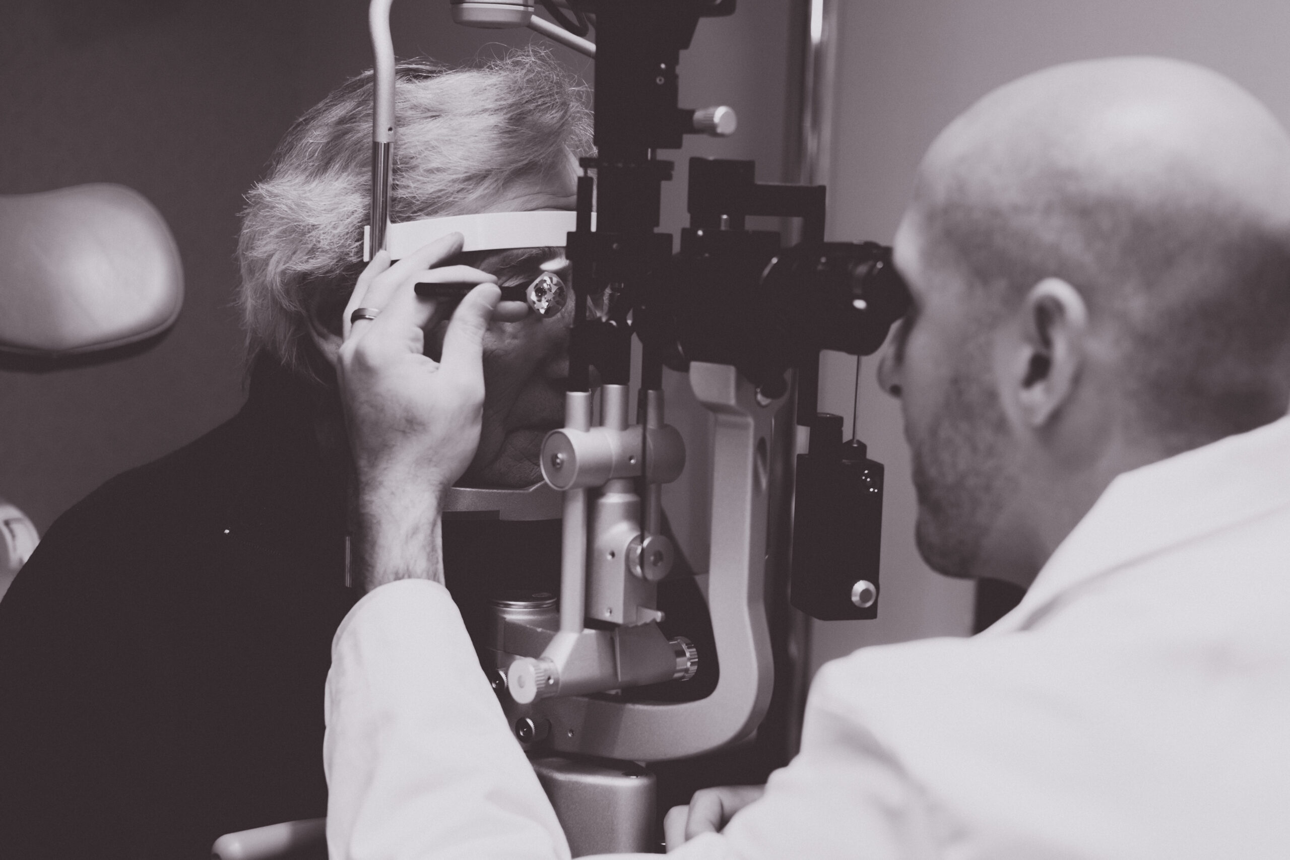
Tonometry measures the pressure within the eye in millimeters of mercury.
The cornea (front of the eye) is gently touched with a very sensitive instrument called an applanator.
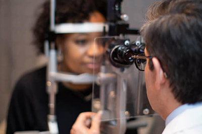
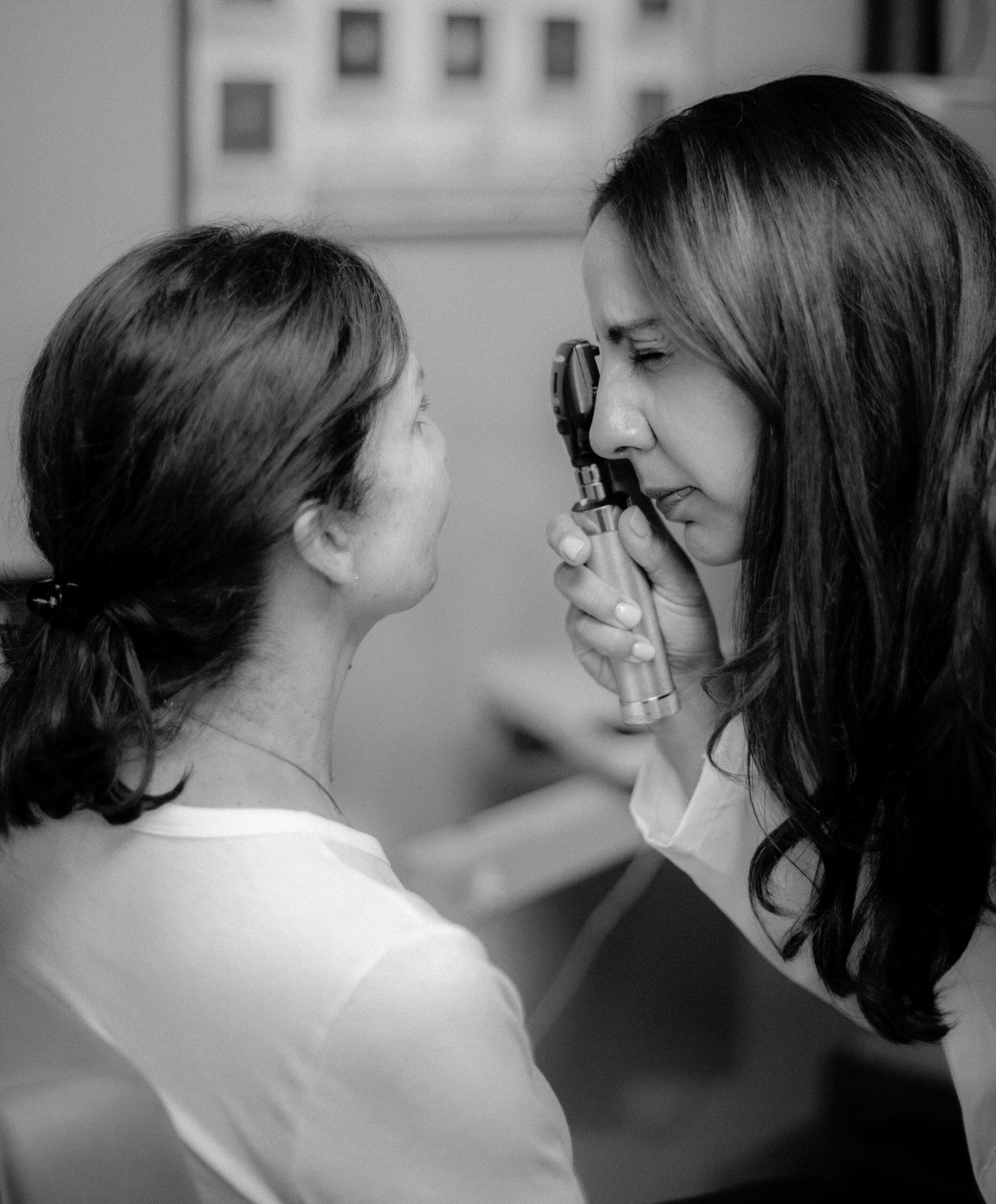
The evaluation of the optic nerve to locate characteristic nerve damage is performed with this instrument.
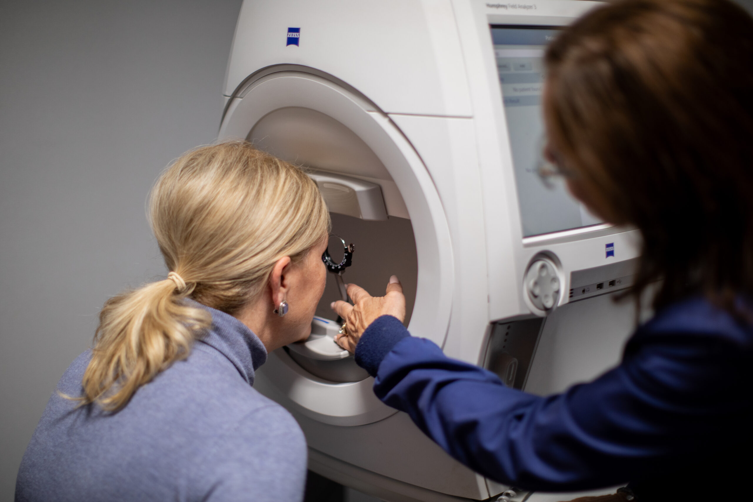
Visual field testing detects and measures the severity of the vision loss due to glaucoma.
This instrument measures the loss of side or peripheral vision, which frequently occurs without the patient’s knowledge.
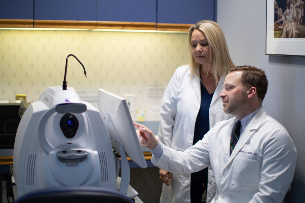
This test uses light waves to take cross-section pictures of your optic nerve and macula. This allows us to see evaluate the nerve fiber layer and cells of your macula (ganglion cells). These measurement’s help with diagnosis of glaucoma and allow us to track one’s stability over time.
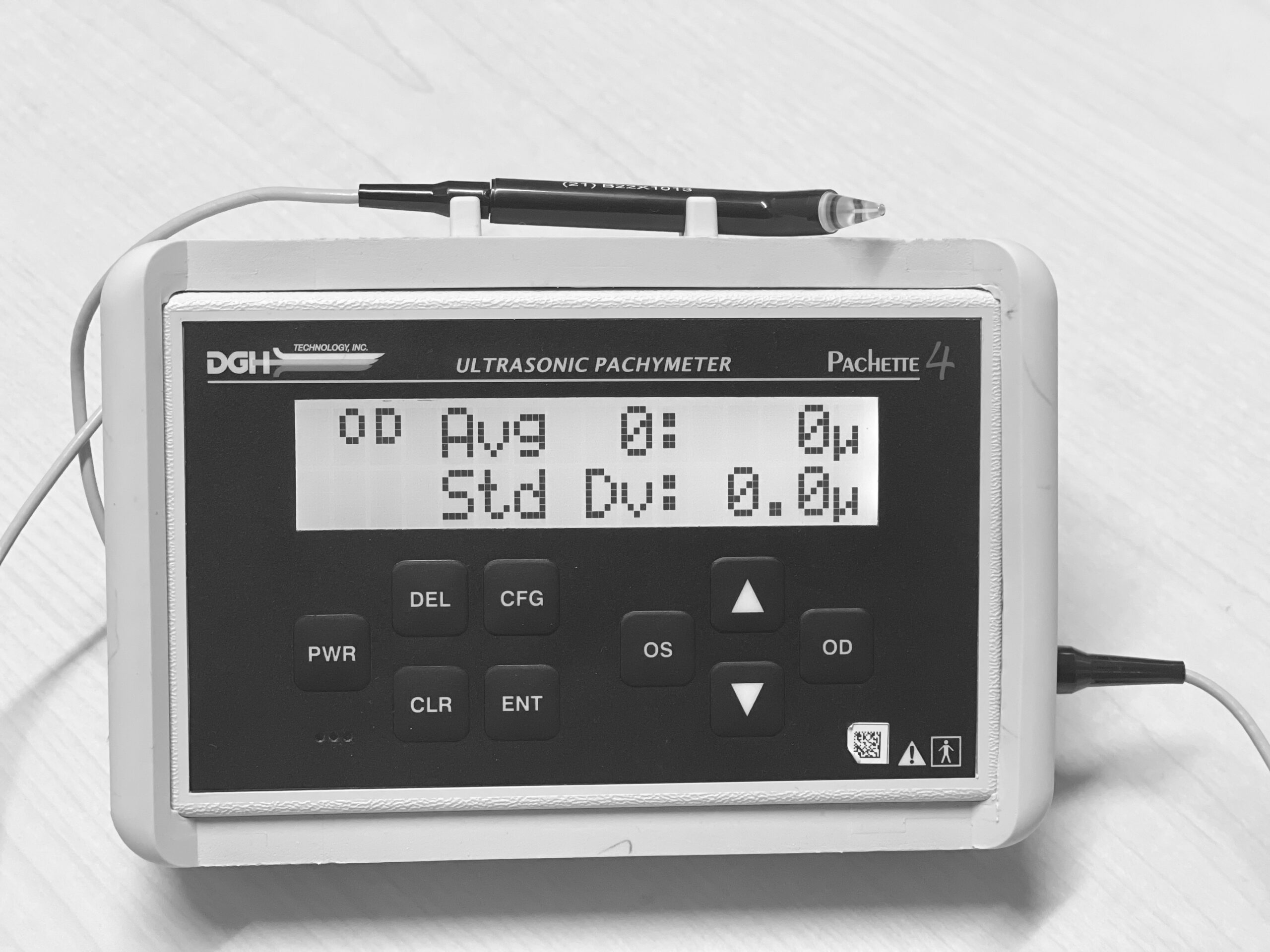
This test measures the thickness of the cornea. Checking the intraocular pressure by tonometry assumes a normal corneal thickness.
If the cornea is too thick or too thin there is an adjustment made in the intraocular pressure based on the pachymetry reading.
This test measures the flexibility of the cornea and sclera (whites of the eye). Checking the intraocular pressure by tonometry assumes a normal corneal hysteresis.
If the corneal hysteresis is low, it may mean that your eye may be more sensitive to elevations or variability in the eye pressure. This can help your doctor more fully assess your risk of developing glaucoma or risk of progression.